Wenn Sie das Fenster schließen, wird Ihre Konfiguration nicht gespeichert, es sei denn, Sie haben Ihren Artikel in die Bestellung aufgenommen oder zu Ihren Favoriten hinzugefügt.
Klicken Sie auf OK, um das MILLIPLEX® MAP-Tool zu schließen oder auf Abbrechen, um zu Ihrer Auswahl zurückzukehren.
Wählen Sie konfigurierbare Panels & Premixed-Kits - ODER - Kits für die zelluläre Signaltransduktion & MAPmates™
Konfigurieren Sie Ihre MILLIPLEX® MAP-Kits und lassen sich den Preis anzeigen.
Konfigurierbare Panels & Premixed-Kits
Unser breites Angebot enthält Multiplex-Panels, für die Sie die Analyten auswählen können, die am besten für Ihre Anwendung geeignet sind. Unter einem separaten Register können Sie das Premixed-Cytokin-Format oder ein Singleplex-Kit wählen.
Kits für die zelluläre Signaltransduktion & MAPmates™
Wählen Sie gebrauchsfertige Kits zur Erforschung gesamter Signalwege oder Prozesse. Oder konfigurieren Sie Ihre eigenen Kits mit Singleplex MAPmates™.
Die folgenden MAPmates™ sollten nicht zusammen analysiert werden: -MAPmates™, die einen unterschiedlichen Assaypuffer erfordern. -Phosphospezifische und MAPmate™ Gesamtkombinationen wie Gesamt-GSK3β und Gesamt-GSK3β (Ser 9). -PanTyr und locusspezifische MAPmates™, z.B. Phospho-EGF-Rezeptor und Phospho-STAT1 (Tyr701). -Mehr als 1 Phospho-MAPmate™ für ein einziges Target (Akt, STAT3). -GAPDH und β-Tubulin können nicht mit Kits oder MAPmates™, die panTyr enthalten, analysiert werden.
.
Bestellnummer
Bestellinformationen
St./Pkg.
Liste
Dieser Artikel wurde zu Ihren Favoriten hinzugefügt.
Wählen Sie bitte Spezies, Panelart, Kit oder Probenart
Um Ihr MILLIPLEX® MAP-Kit zu konfigurieren, wählen Sie zunächst eine Spezies, eine Panelart und/oder ein Kit.
Custom Premix Selecting "Custom Premix" option means that all of the beads you have chosen will be premixed in manufacturing before the kit is sent to you.
Catalogue Number
Ordering Description
Qty/Pack
List
Dieser Artikel wurde zu Ihren Favoriten hinzugefügt.
Spezies
Panelart
Gewähltes Kit
Menge
Bestellnummer
Bestellinformationen
St./Pkg.
Listenpreis
96-Well Plate
Menge
Bestellnummer
Bestellinformationen
St./Pkg.
Listenpreis
Weitere Reagenzien hinzufügen (MAPmates erfordern die Verwendung eines Puffer- und Detektionskits)
Menge
Bestellnummer
Bestellinformationen
St./Pkg.
Listenpreis
48-602MAG
Buffer Detection Kit for Magnetic Beads
1 Kit
Platzsparende Option Kunden, die mehrere Kits kaufen, können ihre Multiplex-Assaykomponenten in Kunststoffbeuteln anstelle von Packungen erhalten, um eine kompaktere Lagerung zu ermöglichen.
Dieser Artikel wurde zu Ihren Favoriten hinzugefügt.
Das Produkt wurde in Ihre Bestellung aufgenommen
Sie können nun ein weiteres Kit konfigurieren, ein Premixed-Kit wählen, zur Kasse gehen oder das Bestell-Tool schließen.
Anti-LGMN/AEP, clone 6E3, Cat. No. MABN2304, is a mouse monoclonal antibody that detects Legumain and is tested for use in ELISA, Immunocytochemistry, Immunohistochemistry (Paraffin), Immunoprecipitation, and Western Blotting.
More>>Anti-LGMN/AEP, clone 6E3, Cat. No. MABN2304, is a mouse monoclonal antibody that detects Legumain and is tested for use in ELISA, Immunocytochemistry, Immunohistochemistry (Paraffin), Immunoprecipitation, and Western Blotting. Less<<
Legumain (UniProt: Q99538; also known as EC: 3.4.22.34, Asparaginyl endopeptidase, AEP, Protease cysteine 1, Delta Secretase) is encoded by the LGMN (also known as PRSC1) gene (Gene ID: 5641) in human. AEP is a lysosomal endopeptidase that displays strict specificity for the hydrolysis of asparaginyl bonds and can simultaneously cleave both amyloid precursor protein (APP) and tau. It cleaves aspartyl bonds slowly, especially under acidic conditions. AEP is synthesized with a signal peptide (aa 1-17) and a propeptide (aa 324-433) that are cleaved off in the mature form. It exists a homodimer before removal of the propeptide and after autocatalytic processing exists as a monomer. In its zymogen form, the uncleaved propeptide blocks access to the active site. AEP is phosphorylated on serine 226 by serine-arginine protein kinase 2 (SRPK2), which accelerates its autocatalytic cleavage and enhances its enzymatic activity. The active form translocates to cytoplasm. In Alzheimer s disease brains AEP is reported to be highly phosphorylated. Depletion of AEP is shown to reduce amyloid peptide production and senile plaque formation in 5XFAD mouse brain that leads to restoration of synaptic activity and cognitive function. Three isoforms of AEP have been described that are produced by alternative splicing. (Ref.: Wang Z-H., et al. (2017). Mol. Cell 67(5); 812-825; Zhang, Z et al (2015). Nat. Commun. 6; 9762).
References
Product Information
Format
Purified
Presentation
Purified mouse monoclonal antibody IgG1 in buffer containing 0.1 M Tris-Glycine (pH 7.4), 150 mM NaCl with 0.05% sodium azide.
Anti-LGMN/AEP, clone 6E3, Cat. No. MABN2304, is a mouse monoclonal antibody that detects Legumain and is tested for use in ELISA, Immunocytochemistry, Immunohistochemistry (Paraffin), Immunoprecipitation, and Western Blotting.
Key Applications
ELISA
Immunocytochemistry
Immunohistochemistry (Paraffin)
Immunoprecipitation
Western Blotting
Application Notes
Tested Applications
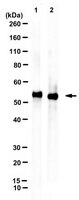
Western Blotting Analysis: A 1:1,000 dilution from a representative lot detected Legumain/AEP in mouse brain tissue lysate.
ELISA Analysis: A representative lot detected Legumain/AEP in ELISA applications (Mattock, K.L., et al. (2010). Atherosclerosis.208(1):83-9; Kang, S.S., et al. (2018). EMBO J. 37(12):e98878).
Immunoprecipitation Analysis: A representative lot immunoprecipitated legumain/AEP in Immunoprecipitation applications (Zhang, Z., et al. (2015). Nat Commun. 6:8762).
Immunohistochemistry (Paraffin) Analysis: A 1:50 dilution from a representative lot detected Legumain/AEP in human placenta tissue sections.
Immunocytochemistry Analysis: A representative lot detected Legumain/AEP in Immunocytochemistry applications (Li, D.N., et al. (2003). J. Biol. Chem. 278(40):38980-90; Wang, Z-H., et al. (2019). Cell Rep. 28(3):655-669).
Western Blotting Analysis: A representative lot detected Legumain/AEP in Western Blotting applications (Probst-Kepper, M., et al. (2009). J Cell Mol Med.;13(9B):3343-57; Zhang, Z., et al. (2015). Nat Commun.;6:8762; Wang, Z-H., et al. (2017). Mol Cell.;67(5):812-825; Zhang, Z., et al. (2017). Nat Struct Mol Biol.;24(8):632-642; Kang, S.S., et al. (2018). EMBO J.;37(12):e98878; Wang, Z-H., et al. (2018). Nat Commun.;9(1):1784; Wang, Z-H., et al. (2018). JCI Insight.;3(16):e99007; Wang, Z-H., et al. (2019). Cell Rep.;28(3):655-669).
Note: Actual optimal working dilutions must be determined by end user as specimens, and experimental conditions may vary with the end user
Biological Information
Immunogen
His-tagged full-length recombinant human legumain.
Epitope
Unknown
Clone
6E3
Concentration
1.0 mg/mL. Please refer to guidance on suggested starting dilutions and/or titers per application and sample type.
Host
Mouse
Specificity
Clone 6E3 is a mouse monoclonal antibody that specifically detects Legumain (AEP).
~55 kDa observed; 49.41 kDa calculated. Uncharacterized bands may be observed in some lysate(s).
Physicochemical Information
Dimensions
Materials Information
Toxicological Information
Safety Information according to GHS
Safety Information
Product Usage Statements
Quality Assurance
Evaluated by Western Blotting in SH-SY5Y cell lysate.
Western Blotting Analysis: A 1:1000 dilution of this antibody detected Legumain/AEP in SH-SY5Y cell lysate.
Usage Statement
Unless otherwise stated in our catalog or other company documentation accompanying the product(s), our products are intended for research use only and are not to be used for any other purpose, which includes but is not limited to, unauthorized commercial uses, in vitro diagnostic uses, ex vivo or in vivo therapeutic uses or any type of consumption or application to humans or animals.
Storage and Shipping Information
Storage Conditions
Recommend storage at +2°C to +8°C. For long term storage antibodies can be kept at -20°C. Avoid repeated freeze-thaws.