Wenn Sie das Fenster schließen, wird Ihre Konfiguration nicht gespeichert, es sei denn, Sie haben Ihren Artikel in die Bestellung aufgenommen oder zu Ihren Favoriten hinzugefügt.
Klicken Sie auf OK, um das MILLIPLEX® MAP-Tool zu schließen oder auf Abbrechen, um zu Ihrer Auswahl zurückzukehren.
Wählen Sie konfigurierbare Panels & Premixed-Kits - ODER - Kits für die zelluläre Signaltransduktion & MAPmates™
Konfigurieren Sie Ihre MILLIPLEX® MAP-Kits und lassen sich den Preis anzeigen.
Konfigurierbare Panels & Premixed-Kits
Unser breites Angebot enthält Multiplex-Panels, für die Sie die Analyten auswählen können, die am besten für Ihre Anwendung geeignet sind. Unter einem separaten Register können Sie das Premixed-Cytokin-Format oder ein Singleplex-Kit wählen.
Kits für die zelluläre Signaltransduktion & MAPmates™
Wählen Sie gebrauchsfertige Kits zur Erforschung gesamter Signalwege oder Prozesse. Oder konfigurieren Sie Ihre eigenen Kits mit Singleplex MAPmates™.
Die folgenden MAPmates™ sollten nicht zusammen analysiert werden: -MAPmates™, die einen unterschiedlichen Assaypuffer erfordern. -Phosphospezifische und MAPmate™ Gesamtkombinationen wie Gesamt-GSK3β und Gesamt-GSK3β (Ser 9). -PanTyr und locusspezifische MAPmates™, z.B. Phospho-EGF-Rezeptor und Phospho-STAT1 (Tyr701). -Mehr als 1 Phospho-MAPmate™ für ein einziges Target (Akt, STAT3). -GAPDH und β-Tubulin können nicht mit Kits oder MAPmates™, die panTyr enthalten, analysiert werden.
.
Bestellnummer
Bestellinformationen
St./Pkg.
Liste
Dieser Artikel wurde zu Ihren Favoriten hinzugefügt.
Wählen Sie bitte Spezies, Panelart, Kit oder Probenart
Um Ihr MILLIPLEX® MAP-Kit zu konfigurieren, wählen Sie zunächst eine Spezies, eine Panelart und/oder ein Kit.
Custom Premix Selecting "Custom Premix" option means that all of the beads you have chosen will be premixed in manufacturing before the kit is sent to you.
Catalogue Number
Ordering Description
Qty/Pack
List
Dieser Artikel wurde zu Ihren Favoriten hinzugefügt.
Spezies
Panelart
Gewähltes Kit
Menge
Bestellnummer
Bestellinformationen
St./Pkg.
Listenpreis
96-Well Plate
Menge
Bestellnummer
Bestellinformationen
St./Pkg.
Listenpreis
Weitere Reagenzien hinzufügen (MAPmates erfordern die Verwendung eines Puffer- und Detektionskits)
Menge
Bestellnummer
Bestellinformationen
St./Pkg.
Listenpreis
48-602MAG
Buffer Detection Kit for Magnetic Beads
1 Kit
Platzsparende Option Kunden, die mehrere Kits kaufen, können ihre Multiplex-Assaykomponenten in Kunststoffbeuteln anstelle von Packungen erhalten, um eine kompaktere Lagerung zu ermöglichen.
Dieser Artikel wurde zu Ihren Favoriten hinzugefügt.
Das Produkt wurde in Ihre Bestellung aufgenommen
Sie können nun ein weiteres Kit konfigurieren, ein Premixed-Kit wählen, zur Kasse gehen oder das Bestell-Tool schließen.
This Anti-Interferon Lambda 4 antibody, clone 4G1 is validated for use in ELISA, Flow Cytometry, Immunocytochemistry, Immunohistochemistry, and Western Blotting for the detection of Interferon Lambda 4.
More>>This Anti-Interferon Lambda 4 antibody, clone 4G1 is validated for use in ELISA, Flow Cytometry, Immunocytochemistry, Immunohistochemistry, and Western Blotting for the detection of Interferon Lambda 4. Less<<
Anti-Interferon Lambda 4, clone 4G1 Antibody: SDB (Sicherheitsdatenblätter), Analysenzertifikate und Qualitätszertifikate, Dossiers, Broschüren und andere verfügbare Dokumente.
Interferon Lambda 4, also known as IFN-lambda-4, and encoded by the gene IFNL4, is an important cytokine that forms part of our innate immunity against viruses. Interferon lambda-4 activates the JAK-STAT signaling pathway and ultimately up regulates several interferon specific stimulated genes. Interferon lambda-4 expression can be stimulated by nucleotide strands particularly ployinosinic-polycytidylic acid (polyI:C) which mimics the double-stranded hepatitis C virus. A genetic variation in the Interferon lambda-4 gene is associated with susceptibility to HCV infection. The mutation of a single base insertion results in gene inactivation resulting in defective immune response to HCV. Interferon lambda-4 signaling operates through Interferon lambda R1 and IL-10R2 receptors and the mutation is associated with defective glycosylation which is required for secretion, thus the amplification of the signal becomes compromised resulting in susceptibility to HCV as well as coronaviruses.
References
Product Information
Format
Purified
Presentation
Purified mouse IgG1κ in buffer containing 0.1 M Tris-Glycine (pH 7.4), 150 mM NaCl with 0.05% sodium azide.
This Anti-Interferon Lambda 4 antibody, clone 4G1 is validated for use in ELISA, Flow Cytometry, Immunocytochemistry, Immunohistochemistry, and Western Blotting for the detection of Interferon Lambda 4.
Key Applications
ELISA
Flow Cytometry
Immunocytochemistry
Immunohistochemistry
Western Blotting
Application Notes
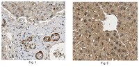
Immunohistochemistry Analysis: A 1:50 dilution from a representative lot detected Interferon Lambda 4 in human and rat liver tissue sections.
ELISA Analysis: A representative lot was conjugated with a sulfo-tag (MSD) and employed as the detection antibody in a sandwich ELISA application. An exogenously expressed IFNL4 HaloTag® fusion was found secreted in the culture media from IFNL4-Halo construct-transfected, but not mock-transfected HepG2 cells (Onabajo, O.O., et al. (2015). J. Interferon. Cytokine Res. 35(11):888-900).
Flow Cytometry Analysis: A representative lot immunostained HepG2 cells transfected to express HaloTag® fusion IFN-λ4 orthologs of elephant, human and non-human primate (chimpanzee, orangutan, rhesus, marmoset, cynomolgus) origins, but not cells expressing IFNL4 of bovine, bat, canine, panda, and porcine species (Paquin, A., et al. (2016). J. Interferon Cytokine Res. 36(1):30-36).
Immunocytochemistry Analysis: Representative lots immunostained HepG2 cells transfected to express HaloTag® fusion of IFN-λ4 orthologs of elephant, human and non-human primate (chimpanzee, orangutan, rhesus, marmoset, cynomolgus) origins (Paquin, A., et al. (2016). J. Interferon Cytokine Res. 36(1):30-36; Prokunina-Olsson, L., et al. (2013). Nat. Genet. 45(2):164-171).
Immunocytochemistry Analysis: A representative lot detected a time-dependent induction of IFNL4 expression in hepatocytes from a donor heterozygous for ss469415590 (TT/ΔG) upon PolyI:C treatment or hepatitis C virus (HCV) strain JFH1HCV infection in cultures (Prokunina-Olsson, L., et al. (2013). Nat. Genet. 45(2):164-171).
Western Blotting Analysis: A representative lot detected HaloTag® fusion IFN-λ4 orthologs of elephant, human and non-human primate (chimpanzee, orangutan, rhesus, marmoset, cynomolgus) origins, but not IFNL4 of bovine, bat, canine, panda, and porcine species transiently expressed in HepG2 cells (Paquin, A., et al. (2016). J. Interferon Cytokine Res. 36(1):30-36).
Western Blotting Analysis: A representative lot detected exogenously expressed IFNL4 HaloTag® fusion constructs in transfected COS-7 and HepG2 cells, but not in untransfected cells. Clone 4G1 does not cross-react with IFN-alpha or IL-28B (Prokunina-Olsson, L., et al. (2013). Nat. Genet. 45(2):164-171).
Biological Information
Immunogen
Linear peptide corresponding to an internal sequence from the N-termninal half of human interferon lambda 4.
Epitope
Internal (N-terminal half).
Clone
4G1
Concentration
1 mg/mL
Host
Mouse
Specificity
Clone 4G1 detected IFNL4 expression induction in primary hepatocytes (e.g. by PolyIC or by HCV) and exogenously overexpressed IFNL4 in HepG2 cells. IFNL4 expression is not detectable in normal liver cells (Dr. Ludmila Prokunina-Olsson, National Cancer Institute, NIH, Bethesda, MD). Clone 4G1 does not cross-react with IFN-alpha or IL-28B (Prokunina-Olsson, L., et al. (2013). Nat. Genet. 45(2):164-171).
Isotype
IgG1κ
Species Reactivity
Human
Non-human Primate
Species Reactivity Note
Human, Non-Human Primate. Not Bovine, Bat, Canine, Panda, or Porcine.
Evaluated by Western Blotting of human IFNL4 recombinant protein.
Western Blotting Analysis: 1.0 µg/mL of this antibody detected 0.1 µg of human IFNL4 recombinant protein.
Usage Statement
Unless otherwise stated in our catalog or other company documentation accompanying the product(s), our products are intended for research use only and are not to be used for any other purpose, which includes but is not limited to, unauthorized commercial uses, in vitro diagnostic uses, ex vivo or in vivo therapeutic uses or any type of consumption or application to humans or animals.
A variant upstream of IFNL3 (IL28B) creating a new interferon gene IFNL4 is associated with impaired clearance of hepatitis C virus. Prokunina-Olsson, Ludmila, et al. Nat. Genet., 45: 164-71 (2013)
2013
Chronic infection with hepatitis C virus (HCV) is a common cause of liver cirrhosis and cancer. We performed RNA sequencing in primary human hepatocytes activated with synthetic double-stranded RNA to mimic HCV infection. Upstream of IFNL3 (IL28B) on chromosome 19q13.13, we discovered a new transiently induced region that harbors a dinucleotide variant ss469415590 (TT or ΔG), which is in high linkage disequilibrium with rs12979860, a genetic marker strongly associated with HCV clearance. ss469415590[ΔG] is a frameshift variant that creates a novel gene, designated IFNL4, encoding the interferon-λ4 protein (IFNL4), which is moderately similar to IFNL3. Compared to rs12979860, ss469415590 is more strongly associated with HCV clearance in individuals of African ancestry, although it provides comparable information in Europeans and Asians. Transient overexpression of IFNL4 in a hepatoma cell line induced STAT1 and STAT2 phosphorylation and the expression of interferon-stimulated genes. Our findings provide new insights into the genetic regulation of HCV clearance and its clinical management.