Wenn Sie das Fenster schließen, wird Ihre Konfiguration nicht gespeichert, es sei denn, Sie haben Ihren Artikel in die Bestellung aufgenommen oder zu Ihren Favoriten hinzugefügt.
Klicken Sie auf OK, um das MILLIPLEX® MAP-Tool zu schließen oder auf Abbrechen, um zu Ihrer Auswahl zurückzukehren.
Wählen Sie konfigurierbare Panels & Premixed-Kits - ODER - Kits für die zelluläre Signaltransduktion & MAPmates™
Konfigurieren Sie Ihre MILLIPLEX® MAP-Kits und lassen sich den Preis anzeigen.
Konfigurierbare Panels & Premixed-Kits
Unser breites Angebot enthält Multiplex-Panels, für die Sie die Analyten auswählen können, die am besten für Ihre Anwendung geeignet sind. Unter einem separaten Register können Sie das Premixed-Cytokin-Format oder ein Singleplex-Kit wählen.
Kits für die zelluläre Signaltransduktion & MAPmates™
Wählen Sie gebrauchsfertige Kits zur Erforschung gesamter Signalwege oder Prozesse. Oder konfigurieren Sie Ihre eigenen Kits mit Singleplex MAPmates™.
Die folgenden MAPmates™ sollten nicht zusammen analysiert werden: -MAPmates™, die einen unterschiedlichen Assaypuffer erfordern. -Phosphospezifische und MAPmate™ Gesamtkombinationen wie Gesamt-GSK3β und Gesamt-GSK3β (Ser 9). -PanTyr und locusspezifische MAPmates™, z.B. Phospho-EGF-Rezeptor und Phospho-STAT1 (Tyr701). -Mehr als 1 Phospho-MAPmate™ für ein einziges Target (Akt, STAT3). -GAPDH und β-Tubulin können nicht mit Kits oder MAPmates™, die panTyr enthalten, analysiert werden.
.
Bestellnummer
Bestellinformationen
St./Pkg.
Liste
Dieser Artikel wurde zu Ihren Favoriten hinzugefügt.
Wählen Sie bitte Spezies, Panelart, Kit oder Probenart
Um Ihr MILLIPLEX® MAP-Kit zu konfigurieren, wählen Sie zunächst eine Spezies, eine Panelart und/oder ein Kit.
Custom Premix Selecting "Custom Premix" option means that all of the beads you have chosen will be premixed in manufacturing before the kit is sent to you.
Catalogue Number
Ordering Description
Qty/Pack
List
Dieser Artikel wurde zu Ihren Favoriten hinzugefügt.
Spezies
Panelart
Gewähltes Kit
Menge
Bestellnummer
Bestellinformationen
St./Pkg.
Listenpreis
96-Well Plate
Menge
Bestellnummer
Bestellinformationen
St./Pkg.
Listenpreis
Weitere Reagenzien hinzufügen (MAPmates erfordern die Verwendung eines Puffer- und Detektionskits)
Menge
Bestellnummer
Bestellinformationen
St./Pkg.
Listenpreis
48-602MAG
Buffer Detection Kit for Magnetic Beads
1 Kit
Platzsparende Option Kunden, die mehrere Kits kaufen, können ihre Multiplex-Assaykomponenten in Kunststoffbeuteln anstelle von Packungen erhalten, um eine kompaktere Lagerung zu ermöglichen.
Dieser Artikel wurde zu Ihren Favoriten hinzugefügt.
Das Produkt wurde in Ihre Bestellung aufgenommen
Sie können nun ein weiteres Kit konfigurieren, ein Premixed-Kit wählen, zur Kasse gehen oder das Bestell-Tool schließen.
Anti-IA-2 antibody, clone 76F is an antibody against IA-2 for use in western blotting, IHC & IP.
More>>Anti-IA-2 antibody, clone 76F is an antibody against IA-2 for use in western blotting, IHC & IP. Less<<
Anti-IA-2, clone 76F Antibody: SDB (Sicherheitsdatenblätter), Analysenzertifikate und Qualitätszertifikate, Dossiers, Broschüren und andere verfügbare Dokumente.
ICA 512, also known as Receptor-type tyrosine-protein phosphatase-like N (R-PTP-N), or Islet cell antigen 512 (ICA 512) or Islet cell autoantigen 3, or PTPIA-2, or IA-2, and encoded by the gene PTPRN/ICA3/ICA512, is a required protein for endocrine and neuroendocrine secretory processes. ICA 512 may be involved in processes specific for neurosecretory granules, such as their biogenesis, trafficking and regulated exocytosis, or ICA 512 may have a more general role in neuroendocrine functions. ICA 512 interacts with phosphorylated syntrophin beta 2/Distrophin, protecting it from protein cleavage by calpain I. Dephosphorylation of Distrophin upon insulin stimulation disrupts the interaction and results in its cleavage. ICA 512 is a membrane protein and found within neuroendocrine secretory granules and endocrine tissues. ICA 512 is expressed in brain, pituitary and gut tissues including the pancreas. ICA 512 is also now used as a diabetes-specific islet autoantigen biomarker in type diabetes patients.
References
Product Information
Format
Purified
Presentation
Purified mouse monoclonal IgG2bκ in buffer containing 0.1 M Tris-Glycine (pH 7.4), 150 mM NaCl with 0.05% sodium azide.
Anti-IA-2 antibody, clone 76F is an antibody against IA-2 for use in western blotting, IHC & IP.
Key Applications
Western Blotting
Immunohistochemistry
Immunoprecipitation
Application Notes
Immunohistochemistry Analysis: A representative lot from an independent laboratory detected IA-2 in rat pancreas tissues and BTC3 cells (Piquer, S., et al. (2006). J Autoimmun. 26(3):215-222.).
Immunoprecipitation Analysis: A representative lot from an independent laboratory immunoprecipitated IA-2 from recombinant rat, mouse, and human IA-2 (Piquer, S., et al. (2006). J Autoimmun. 26(3):215-222.).
Biological Information
Immunogen
His-tagged recombinant protein corresponding to the residues 626-630 found in juxtamembrane region of human and mouse IA-2, but not IA-2b of human IA-2.
Epitope
This antibody recognizes residues 626-630 found in juxtamembrane region of human and mouse IA-2, but not in IA-2b.
Clone
76F
Concentration
Please refer to the Certificate of Analysis for the lot-specific concentration.
~65 kDa observed. The full length protein is cleaved to yield a 65 kDa mature protein form that contains a lumenal domain, and an intracellular cytoplasmic domain characterized by a juxtamembrane region (Piquer, S., et al. (2006). J Autoimmun. 26(3):215-222.).
Physicochemical Information
Dimensions
Materials Information
Toxicological Information
Safety Information according to GHS
Safety Information
Product Usage Statements
Quality Assurance
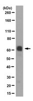
Evaluated by Western Blotting in human pancreas tissue lysate.
Western Blotting Analysis: 0.5 µg/mL of this antibody detected IA-2 in 10 µg of human pancreas tissue lysate.
Usage Statement
Unless otherwise stated in our catalog or other company documentation accompanying the product(s), our products are intended for research use only and are not to be used for any other purpose, which includes but is not limited to, unauthorized commercial uses, in vitro diagnostic uses, ex vivo or in vivo therapeutic uses or any type of consumption or application to humans or animals.
Monoclonal antibody 76F distinguishes IA-2 from IA-2beta and overlaps an autoantibody epitope. Piquer, Sandra, et al. J. Autoimmun., 26: 215-22 (2006)
2005
IA-2 and IA-2beta are highly related proteins that are autoantigens in type 1 diabetes, and provide a model for developing reagents and assays that distinguish similar proteins with unique autoantibody epitopes. Monoclonal antibodies (mAb) to IA-2 and IA-2beta were prepared and tested for their ability to bind to the related proteins and their ability to compete for specific autoantibody epitope binding by sera from patients with type 1 diabetes. Monoclonal antibodies that specifically bound IA-2 (76F) or bound both IA-2 and IA-2beta (A9) were isolated and characterized. 76F mAb recognized IA-2 of human, rat and mouse origin in native and denatured forms and had an epitope specificity for residues 626-630 (FEYQD) which are found in the juxtamembrane (JM) region of human and mouse IA-2, but not IA-2beta. This region overlaps with the autoantibody epitope JM2. Binding to the 76F monoclonal antibody was specifically inhibited by sera with antibodies to the JM2 epitope but not with antibodies to the adjacent JM1 epitope, indicating that unique epitopes can be distinguished by this approach. 76F mAb has the unique property to distinguish between the two closely related autoantigens IA-2 and IA-2beta by targeting an IA-2 specific epitope of the juxtamembrane region. The findings define an approach to develop assays for specific antibody epitope measurements which may be relevant for disease prognosis and monitoring intervention therapies.