Wenn Sie das Fenster schließen, wird Ihre Konfiguration nicht gespeichert, es sei denn, Sie haben Ihren Artikel in die Bestellung aufgenommen oder zu Ihren Favoriten hinzugefügt.
Klicken Sie auf OK, um das MILLIPLEX® MAP-Tool zu schließen oder auf Abbrechen, um zu Ihrer Auswahl zurückzukehren.
Wählen Sie konfigurierbare Panels & Premixed-Kits - ODER - Kits für die zelluläre Signaltransduktion & MAPmates™
Konfigurieren Sie Ihre MILLIPLEX® MAP-Kits und lassen sich den Preis anzeigen.
Konfigurierbare Panels & Premixed-Kits
Unser breites Angebot enthält Multiplex-Panels, für die Sie die Analyten auswählen können, die am besten für Ihre Anwendung geeignet sind. Unter einem separaten Register können Sie das Premixed-Cytokin-Format oder ein Singleplex-Kit wählen.
Kits für die zelluläre Signaltransduktion & MAPmates™
Wählen Sie gebrauchsfertige Kits zur Erforschung gesamter Signalwege oder Prozesse. Oder konfigurieren Sie Ihre eigenen Kits mit Singleplex MAPmates™.
Die folgenden MAPmates™ sollten nicht zusammen analysiert werden: -MAPmates™, die einen unterschiedlichen Assaypuffer erfordern. -Phosphospezifische und MAPmate™ Gesamtkombinationen wie Gesamt-GSK3β und Gesamt-GSK3β (Ser 9). -PanTyr und locusspezifische MAPmates™, z.B. Phospho-EGF-Rezeptor und Phospho-STAT1 (Tyr701). -Mehr als 1 Phospho-MAPmate™ für ein einziges Target (Akt, STAT3). -GAPDH und β-Tubulin können nicht mit Kits oder MAPmates™, die panTyr enthalten, analysiert werden.
.
Bestellnummer
Bestellinformationen
St./Pkg.
Liste
Dieser Artikel wurde zu Ihren Favoriten hinzugefügt.
Wählen Sie bitte Spezies, Panelart, Kit oder Probenart
Um Ihr MILLIPLEX® MAP-Kit zu konfigurieren, wählen Sie zunächst eine Spezies, eine Panelart und/oder ein Kit.
Custom Premix Selecting "Custom Premix" option means that all of the beads you have chosen will be premixed in manufacturing before the kit is sent to you.
Catalogue Number
Ordering Description
Qty/Pack
List
Dieser Artikel wurde zu Ihren Favoriten hinzugefügt.
Spezies
Panelart
Gewähltes Kit
Menge
Bestellnummer
Bestellinformationen
St./Pkg.
Listenpreis
96-Well Plate
Menge
Bestellnummer
Bestellinformationen
St./Pkg.
Listenpreis
Weitere Reagenzien hinzufügen (MAPmates erfordern die Verwendung eines Puffer- und Detektionskits)
Menge
Bestellnummer
Bestellinformationen
St./Pkg.
Listenpreis
48-602MAG
Buffer Detection Kit for Magnetic Beads
1 Kit
Platzsparende Option Kunden, die mehrere Kits kaufen, können ihre Multiplex-Assaykomponenten in Kunststoffbeuteln anstelle von Packungen erhalten, um eine kompaktere Lagerung zu ermöglichen.
Dieser Artikel wurde zu Ihren Favoriten hinzugefügt.
Das Produkt wurde in Ihre Bestellung aufgenommen
Sie können nun ein weiteres Kit konfigurieren, ein Premixed-Kit wählen, zur Kasse gehen oder das Bestell-Tool schließen.
This mouse monoclonal Anti-CLEC-2 Antibody, clone AYP1, Cat. No. MABF957 is validated for use in Flow Cytometry, Function Assay, and Immunoprecipitation for the detection of CLEC-2.
More>>This mouse monoclonal Anti-CLEC-2 Antibody, clone AYP1, Cat. No. MABF957 is validated for use in Flow Cytometry, Function Assay, and Immunoprecipitation for the detection of CLEC-2. Less<<
Anti-CLEC-2 Antibody, clone AYP1 : SDB (Sicherheitsdatenblätter), Analysenzertifikate und Qualitätszertifikate, Dossiers, Broschüren und andere verfügbare Dokumente.
C-type lectin domain family 1 member B (UniProt Q9P126; also known as C-type lectin-like receptor 2, CLEC-2) is encoded by the CLEC1B (also known as CLEC2, UNQ721/PRO1384) gene (Gene ID 51266) in human. CLEC-2 is a type II single-transmembrane (a.a. 34-54) protein with a large extracellular (a.a. 55-229) region that contains a C-type lectin domain (a.a. 109-217) and a short N-terminal cytoplasmic tail (a.a. 1-33) with a single hem-immunoreceptor tyrosine-based activation motif (hemITAM; a.a. 7-10). CLEC-2 expression is restricted to platelets, where it mediates platelets activation via its hemITAM motif, leading to proteolytic cleavage of two other platelet ITAM receptors, glycoprotein (GP)VI and Fc RIIa, but not of CLEC-2 itself. CLEC-2 functions as the receptor for the type I transmembrane GP podoplanin that is widely expressed outside of the vasculature, including lymphatic endothelial cells, type 1 lung alveolar cells, lymph node stromal cells, the choroid plexus epithelium, inflammatory macrophages, as well as a subset of activated T-helper (Th)17 cells. Patients with rheumatoid arthritis show increased plasma levels of CLEC-2-positive microparticles. These microparticles are derived from activated platelets and are negative for GPVI, while microparticles in healthy donors are predominantly derived from megakaryocytes and are positive for both CLEC-2 and GPVI. Platelet-specific deletion of CLEC-2 or one of its downstream signaling proteins, Syk, SLP-76, or PLC 2, leads to a number of developmental problems in mice, including blood-lymphatic mixing in midgestation.
This mouse monoclonal Anti-CLEC-2 Antibody, clone AYP1, Cat. No. MABF957 is validated for use in Flow Cytometry, Function Assay, and Immunoprecipitation for the detection of CLEC-2.
Key Applications
Flow Cytometry
Application Notes
Flow Cytometry Analysis: A representative lot detected doxycycline-dependent expression induction of exogenously transfected CLEC-2/CLEC1B using T-REx 293 cells (Gitz, E., et al. (2014). Blood. 124(14):2262-2270).
Flow Cytometry Analysis: A representative lot (pre-conjugated with Alexa Fluor® 488) detected CLEC-2/CLEC1B expression on the surface of platelets and CD41-positive microparticles in platelet-rich plasma (PRP), but not on monocytes, neutrophils, dendritic cells, B- or T-cells (Gitz, E., et al. (2014). Blood. 124(14):2262-2270).
Function Assay: Clone AYP1 Fab fragment (2.5 µg/mL) cross-linked with anti-mouse Fab-specific F(ab)2 fragments, but not AYP1 Fab or anti-mouse F(ab)2 alone, induced surface P-selectin expression and the aggregation of washed human platelets (Gitz, E., et al. (2014). Blood. 124(14):2262-2270).
Immunoprecipitation Analysis: A representative lot immunoprecipitated CLEC-2/CLEC1B from human platelet lysates. Rhodocytin stimulation prior to cell lysis and IP induced CLEC-2/CLEC1B tyrosine phosphorylation (Gitz, E., et al. (2014). Blood. 124(14):2262-2270).
Biological Information
Immunogen
His-tagged recombinant human CLEC-2 extracellular fragment.
Epitope
extracellular domain
Clone
AYP1
Concentration
Please refer to lot specific datasheet.
Host
Mouse
Specificity
Clone AYP1 targets an extracellular epitope present in both spliced isoforms of human CLEC-2/CLEC1B reported by UniProt (Q9P126).
26.60 kDa (isoform 1) and 23.10 kDa (isoform 2). ~32/40 kDa doublet reported due to differential glycosylation (Gitz, E., et al. (2014). Blood. 124(14):2262-2270).
Physicochemical Information
Dimensions
Materials Information
Toxicological Information
Safety Information according to GHS
Safety Information
Product Usage Statements
Quality Assurance
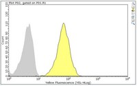
Evaluated by Flow Cytometry in human platelets.
Flow Cytometry Analysis: 0.2 µL of this antibody detected CLEC-2/CLEC1B on the surface of human platelets.
Usage Statement
Unless otherwise stated in our catalog or other company documentation accompanying the product(s), our products are intended for research use only and are not to be used for any other purpose, which includes but is not limited to, unauthorized commercial uses, in vitro diagnostic uses, ex vivo or in vivo therapeutic uses or any type of consumption or application to humans or animals.
Storage and Shipping Information
Storage Conditions
Stable for 1 year at -20°C from date of receipt.
Handling Recommendations: Upon receipt and prior to removing the cap, centrifuge the vial and gently mix the solution. Aliquot into microcentrifuge tubes and store at -20°C. Avoid repeated freeze/thaw cycles, which may damage IgG and affect product performance.