Wenn Sie das Fenster schließen, wird Ihre Konfiguration nicht gespeichert, es sei denn, Sie haben Ihren Artikel in die Bestellung aufgenommen oder zu Ihren Favoriten hinzugefügt.
Klicken Sie auf OK, um das MILLIPLEX® MAP-Tool zu schließen oder auf Abbrechen, um zu Ihrer Auswahl zurückzukehren.
Wählen Sie konfigurierbare Panels & Premixed-Kits - ODER - Kits für die zelluläre Signaltransduktion & MAPmates™
Konfigurieren Sie Ihre MILLIPLEX® MAP-Kits und lassen sich den Preis anzeigen.
Konfigurierbare Panels & Premixed-Kits
Unser breites Angebot enthält Multiplex-Panels, für die Sie die Analyten auswählen können, die am besten für Ihre Anwendung geeignet sind. Unter einem separaten Register können Sie das Premixed-Cytokin-Format oder ein Singleplex-Kit wählen.
Kits für die zelluläre Signaltransduktion & MAPmates™
Wählen Sie gebrauchsfertige Kits zur Erforschung gesamter Signalwege oder Prozesse. Oder konfigurieren Sie Ihre eigenen Kits mit Singleplex MAPmates™.
Die folgenden MAPmates™ sollten nicht zusammen analysiert werden: -MAPmates™, die einen unterschiedlichen Assaypuffer erfordern. -Phosphospezifische und MAPmate™ Gesamtkombinationen wie Gesamt-GSK3β und Gesamt-GSK3β (Ser 9). -PanTyr und locusspezifische MAPmates™, z.B. Phospho-EGF-Rezeptor und Phospho-STAT1 (Tyr701). -Mehr als 1 Phospho-MAPmate™ für ein einziges Target (Akt, STAT3). -GAPDH und β-Tubulin können nicht mit Kits oder MAPmates™, die panTyr enthalten, analysiert werden.
.
Bestellnummer
Bestellinformationen
St./Pkg.
Liste
Dieser Artikel wurde zu Ihren Favoriten hinzugefügt.
Wählen Sie bitte Spezies, Panelart, Kit oder Probenart
Um Ihr MILLIPLEX® MAP-Kit zu konfigurieren, wählen Sie zunächst eine Spezies, eine Panelart und/oder ein Kit.
Custom Premix Selecting "Custom Premix" option means that all of the beads you have chosen will be premixed in manufacturing before the kit is sent to you.
Catalogue Number
Ordering Description
Qty/Pack
List
Dieser Artikel wurde zu Ihren Favoriten hinzugefügt.
Spezies
Panelart
Gewähltes Kit
Menge
Bestellnummer
Bestellinformationen
St./Pkg.
Listenpreis
96-Well Plate
Menge
Bestellnummer
Bestellinformationen
St./Pkg.
Listenpreis
Weitere Reagenzien hinzufügen (MAPmates erfordern die Verwendung eines Puffer- und Detektionskits)
Menge
Bestellnummer
Bestellinformationen
St./Pkg.
Listenpreis
48-602MAG
Buffer Detection Kit for Magnetic Beads
1 Kit
Platzsparende Option Kunden, die mehrere Kits kaufen, können ihre Multiplex-Assaykomponenten in Kunststoffbeuteln anstelle von Packungen erhalten, um eine kompaktere Lagerung zu ermöglichen.
Dieser Artikel wurde zu Ihren Favoriten hinzugefügt.
Das Produkt wurde in Ihre Bestellung aufgenommen
Sie können nun ein weiteres Kit konfigurieren, ein Premixed-Kit wählen, zur Kasse gehen oder das Bestell-Tool schließen.
06-1295
Sigma-AldrichAnti-CDT1 Antibody
Use Anti-CDT1 Antibody (Rabbit Polyclonal Antibody) validated in WB to detect CDT1 also known as DNA replication factor, Double parked homolog.
More>>Use Anti-CDT1 Antibody (Rabbit Polyclonal Antibody) validated in WB to detect CDT1 also known as DNA replication factor, Double parked homolog. Less<<
Anti-CDT1 Antibody: SDB (Sicherheitsdatenblätter), Analysenzertifikate und Qualitätszertifikate, Dossiers, Broschüren und andere verfügbare Dokumente.
CDT1 (Chromatin Licensing and DNA replication Factor 1) is an essential component of the cell cycle machinery. It plays an important role in insuring that there is only one round of DNA replication per cell cycle. CDT1 is a known substrate of Cdk2 and Cdk4 (Liu, E. 2004). It is presented in G1 of the cell cycle and is degraded as cells enter S phase (Liu, E. 2004). In higher eukaryotes, CDT1 is known to interact with Geminin, a protein that is degraded during mitosis, that inhibits it. CDT1 is marked for degradation by two E3 ligases: SCF complex and the CUL4-DDB1 complex (Sansam, 2006).
Use Anti-CDT1 Antibody (Rabbit Polyclonal Antibody) validated in WB to detect CDT1 also known as DNA replication factor, Double parked homolog.
Key Applications
Western Blotting
Application Notes
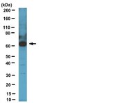
Western Blot Analysis: A 1:1,000 dilution from a previous lot detected CDT1 in C2C12, Hek293, HeLa, HepG2, Huvec, Jurkat, L6, mouse brain, NIH/3T3, PC3, and RAW264.7 cell lysates.
Biological Information
Immunogen
KLH-conjugated linear peptide corresponding to human CDT1.
FUNCTION: Cooperates with CDC6 to promote the loading of the mini-chromosome maintenance complex onto chromatin to form the pre-replication complex necessary to initiate DNA replication. Binds DNA in a sequence-, strand-, and conformation-independent manner. Potential oncogene. SUBUNIT STRUCTURE: Interaction with GMNN inhibits both binding of the MCM complex to origins of replication and DNA-binding activity. SUBCELLULAR LOCATION: Nucleus. DEVELOPMENTAL STAGE: Present during G1 and early S phase of the cell cycle. Degraded during the late S, G2, and M phases. PTM : Ubiquitinated, leading to its subsequent degradation. The interaction with GMNN protects it against ubiquitination. Phosphorylated by cyclin A-dependent kinases which results in the binding of CDT1 to the F-box protein SKP2 and subsequent degradation. Binding to GMNN is not affected by phosphorylation. SEQUENCE SIMILARITIES: Belongs to the Cdt1 family. SEQUENCE CAUTION: The sequence AF070552 differs from that shown. Reason: Frameshift at positions 278 and 312.
Molecular Weight
~60 kDa
Physicochemical Information
Dimensions
Materials Information
Toxicological Information
Safety Information according to GHS
Safety Information
Product Usage Statements
Quality Assurance
Evaluated by Western Blot in HeLa cell lysate. Western Blot Analysis: A 1:1,000 dilution of this antibody detected CDT1 in 10 µg of HeLa cell lysate.
Usage Statement
Unless otherwise stated in our catalog or other company documentation accompanying the product(s), our products are intended for research use only and are not to be used for any other purpose, which includes but is not limited to, unauthorized commercial uses, in vitro diagnostic uses, ex vivo or in vivo therapeutic uses or any type of consumption or application to humans or animals.
Storage and Shipping Information
Storage Conditions
Stable for 1 year at -20°C from date of receipt. Handling Recommendations: Upon first thaw, and prior to removing the cap, centrifuge the vial and gently mix the solution. Aliquot into microcentrifuge tubes and store at -20°C. Avoid repeated freeze/thaw cycles, which may damage IgG and affect product performance.
Infection by the autonomous parvovirus minute virus of mice (MVM) induces a vigorous DNA damage response in host cells which it utilizes for its efficient replication. Although p53 remains activated, p21 protein levels remain low throughout the course of infection. We show here that efficient MVM replication required the targeting for degradation of p21 during this time by the CRL4Cdt2 E3-ubiquitin ligase which became re-localized to MVM replication centers. PCNA provides a molecular platform for substrate recognition by the CRL4Cdt2 E3-ubiquitin ligase and p21 targeting during MVM infection required its interaction both with Cdt2 and PCNA. PCNA is also an important co-factor for MVM replication which can be antagonized by p21 in vitro. Expression of a stable p21 mutant that retained interaction with PCNA inhibited MVM replication, while a stable p21 mutant which lacked this interaction did not. Thus, while interaction with PCNA was important for targeting p21 to the CRL4Cdt2 ligase re-localized to MVM replication centers, efficient viral replication required subsequent depletion of p21 to abrogate its inhibition of PCNA.
Inhibition of NEDD8-activating enzyme (NAE) has emerged as a highly promising approach to treat cancer through the adenosine sulfamate analog MLN4924. Here, we show that selective pressure results in HCT116 colorectal carcinoma cells with decreased MLN4924 sensitivity and identify a single-nucleotide transition that changes alanine 171 to threonine (A171T) of the NAE subunit UBA3. This reduces the enzyme's affinity for MLN4924 and ATP while increasing NEDD8 activation at physiological ATP concentrations. Expression of UBA3 A171T is sufficient to decrease MLN4924 sensitivity of naive HCT116 cells, indicating that it is a dominant suppressor of MLN4924-mediated cell death. Our data suggest that the on-target potency of MLN4924 selects for a point mutation in NAE that overcomes the molecule's inhibitory effects, allowing cancer cell survival.
Minichromosome maintenance helicase paralog MCM9 is dispensible for DNA replication but functions in germ-line stem cells and tumor suppression. Hartford, SA; Luo, Y; Southard, TL; Min, IM; Lis, JT; Schimenti, JC Proceedings of the National Academy of Sciences of the United States of America
108
17702-7
2010
Effective DNA replication is critical to the health and reproductive success of organisms. The six MCM2-7 proteins, which form the replicative helicase, are essential for high-fidelity replication of the genome. Many eukaryotes have a divergent paralog, MCM9, that was reported to be essential for loading MCM2-7 onto replication origins in the Xenopus oocyte extract system. To address the in vivo role of mammalian MCM9, we created and analyzed the phenotypes of mice with various mutations in Mcm9 and an intronic DNA replication-related gene Asf1a. Ablation of Mcm9 was compatible with cell proliferation and mouse viability, showing that it is nonessential for MCM2-7 loading or DNA replication. Mcm9 mutants underwent p53-independent embryonic germ-cell depletion in both sexes, with males also exhibiting defective spermatogonial stem-cell renewal. MCM9-deficient cells had elevated genomic instability and defective cell cycle reentry following replication stress, and mutant animals were prone to sex-specific cancers, most notably hepatocellular carcinoma in males. The phenotypes of mutant mice and cells suggest that MCM9 evolved a specialized but nonessential role in DNA replication or replication-linked quality-control mechanisms that are especially important for germ-line stem cells, and also for tumor suppression and genome maintenance in the soma.