Wenn Sie das Fenster schließen, wird Ihre Konfiguration nicht gespeichert, es sei denn, Sie haben Ihren Artikel in die Bestellung aufgenommen oder zu Ihren Favoriten hinzugefügt.
Klicken Sie auf OK, um das MILLIPLEX® MAP-Tool zu schließen oder auf Abbrechen, um zu Ihrer Auswahl zurückzukehren.
Wählen Sie konfigurierbare Panels & Premixed-Kits - ODER - Kits für die zelluläre Signaltransduktion & MAPmates™
Konfigurieren Sie Ihre MILLIPLEX® MAP-Kits und lassen sich den Preis anzeigen.
Konfigurierbare Panels & Premixed-Kits
Unser breites Angebot enthält Multiplex-Panels, für die Sie die Analyten auswählen können, die am besten für Ihre Anwendung geeignet sind. Unter einem separaten Register können Sie das Premixed-Cytokin-Format oder ein Singleplex-Kit wählen.
Kits für die zelluläre Signaltransduktion & MAPmates™
Wählen Sie gebrauchsfertige Kits zur Erforschung gesamter Signalwege oder Prozesse. Oder konfigurieren Sie Ihre eigenen Kits mit Singleplex MAPmates™.
Die folgenden MAPmates™ sollten nicht zusammen analysiert werden: -MAPmates™, die einen unterschiedlichen Assaypuffer erfordern. -Phosphospezifische und MAPmate™ Gesamtkombinationen wie Gesamt-GSK3β und Gesamt-GSK3β (Ser 9). -PanTyr und locusspezifische MAPmates™, z.B. Phospho-EGF-Rezeptor und Phospho-STAT1 (Tyr701). -Mehr als 1 Phospho-MAPmate™ für ein einziges Target (Akt, STAT3). -GAPDH und β-Tubulin können nicht mit Kits oder MAPmates™, die panTyr enthalten, analysiert werden.
.
Bestellnummer
Bestellinformationen
St./Pkg.
Liste
Dieser Artikel wurde zu Ihren Favoriten hinzugefügt.
Wählen Sie bitte Spezies, Panelart, Kit oder Probenart
Um Ihr MILLIPLEX® MAP-Kit zu konfigurieren, wählen Sie zunächst eine Spezies, eine Panelart und/oder ein Kit.
Custom Premix Selecting "Custom Premix" option means that all of the beads you have chosen will be premixed in manufacturing before the kit is sent to you.
Catalogue Number
Ordering Description
Qty/Pack
List
Dieser Artikel wurde zu Ihren Favoriten hinzugefügt.
Spezies
Panelart
Gewähltes Kit
Menge
Bestellnummer
Bestellinformationen
St./Pkg.
Listenpreis
96-Well Plate
Menge
Bestellnummer
Bestellinformationen
St./Pkg.
Listenpreis
Weitere Reagenzien hinzufügen (MAPmates erfordern die Verwendung eines Puffer- und Detektionskits)
Menge
Bestellnummer
Bestellinformationen
St./Pkg.
Listenpreis
48-602MAG
Buffer Detection Kit for Magnetic Beads
1 Kit
Platzsparende Option Kunden, die mehrere Kits kaufen, können ihre Multiplex-Assaykomponenten in Kunststoffbeuteln anstelle von Packungen erhalten, um eine kompaktere Lagerung zu ermöglichen.
Dieser Artikel wurde zu Ihren Favoriten hinzugefügt.
Das Produkt wurde in Ihre Bestellung aufgenommen
Sie können nun ein weiteres Kit konfigurieren, ein Premixed-Kit wählen, zur Kasse gehen oder das Bestell-Tool schließen.
ABS314
Sigma-AldrichAnti-APPL1 Antibody
Detect Adapter protein containing PH domain, PTB domain & leucine zipper motif 1 using this rabbit polyclonal antibody, Anti-APPL1 Antibody validated for use in western blotting, IP, ICC & IHC.
More>>Detect Adapter protein containing PH domain, PTB domain & leucine zipper motif 1 using this rabbit polyclonal antibody, Anti-APPL1 Antibody validated for use in western blotting, IP, ICC & IHC. Less<<
Anti-APPL1 Antibody: SDB (Sicherheitsdatenblätter), Analysenzertifikate und Qualitätszertifikate, Dossiers, Broschüren und andere verfügbare Dokumente.
Adapter protein containing PH domain, PTB domain and leucine zipper motif 1
Background Information
Adapter protein containing PH domain, PTB domain and leucine zipper motif 1 (APPL1) is also called DCC-interacting protein 13-alpha (Dip13-alpha). APPL1 is necessary for cell proliferation in response to extracellular signals and links Rab5 to nuclear signal transduction. APPL1 is expressed at high levels in heart, ovary, pancreas, and skeletal muscle tissues.
References
Product Information
Format
Affinity Purified
Presentation
Purified rabbit polyclonal in buffer containing PBS with 0.05% sodium azide.
Detect Adapter protein containing PH domain, PTB domain & leucine zipper motif 1 using this rabbit polyclonal antibody, Anti-APPL1 Antibody validated for use in western blotting, IP, ICC & IHC.
Key Applications
Western Blotting
Immunoprecipitation
Immunocytochemistry
Immunohistochemistry
Application Notes
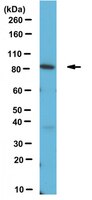
Western Blotting Analysis: 0.5 µg/mL of this antibody detected APPL1 in 10 µg of L6 and NIH/3T3 cell lysate.
Immunoprecipitation Analysis: A representative lot from an independent laboratory immunoprecipitated APPL1 in HUVEC cell lysate (Lin, D. C., et al. (2006). Mol Cell Biol. 26(23):8928-8941.).
Immunohistochemistry Analysis: A representative lot from an independent laboratory detected APPL1 in liver tissues from mice fed with either a standard diet, high fat diet, or a high fat diet with exercise (Marinho, R., et al. (2012). J Cell Physiol. 227(7):2917-2926.).
Immunocytochemistry Analysis: A representative lot from an independent laboratory detected APPL1 in pancreatic islets and impaired GSIS cells of dietary and obese mice (Cheng, K. K., et al. (2009). Cell Metab. 9(5):417-427.).
Biological Information
Immunogen
Recombinant protein corresponding to human APPL1.
Concentration
Please refer to the Certificate of Analysis for the lot-specific concentration.
~84 kDa observed. Uncharacterized band(s) may be observed in some cell lysates.
Physicochemical Information
Dimensions
Materials Information
Toxicological Information
Safety Information according to GHS
Safety Information
Product Usage Statements
Quality Assurance
Evaluated by Western Blotting in A431 cell lysate.
Western Blotting Analysis: 0.5 µg/mL of this antibody detected APPL1 in 10 µg of A431 cell lysate.
Usage Statement
Unless otherwise stated in our catalog or other company documentation accompanying the product(s), our products are intended for research use only and are not to be used for any other purpose, which includes but is not limited to, unauthorized commercial uses, in vitro diagnostic uses, ex vivo or in vivo therapeutic uses or any type of consumption or application to humans or animals.
Endurance exercise training increases APPL1 expression and improves insulin signaling in the hepatic tissue of diet-induced obese mice, independently of weight loss. Marinho, R, et al. J. Cell. Physiol., 227: 2917-26 (2012)
2011
Hepatic insulin resistance is the major contributor to fasting hyperglycemia in type 2 diabetes. The protein kinase Akt plays a central role in the suppression of gluconeogenesis involving forkhead box O1 (Foxo1) and peroxisome proliferator-activated receptor gamma co-activator 1 alpha (PGC-1α), and in the control of glycogen synthesis involving the glycogen synthase kinase beta (GSK3β) in the liver. It has been demonstrated that endosomal adaptor protein APPL1 interacts with Akt and blocks the association of Akt with its endogenous inhibitor, tribbles-related protein 3 (TRB3), improving the action of insulin in the liver. Here, we demonstrated that chronic exercise increased the basal levels and insulin-induced Akt serine phosphorylation in the liver of diet-induced obese mice. Endurance training was able to increase APPL1 expression and the interaction between APPL1 and Akt. Conversely, training reduced both TRB3 expression and TRB3 and Akt association. The positive effects of exercise on insulin action are reinforced by our findings that showed that trained mice presented an increase in Foxo1 phosphorylation and Foxo1/PGC-1α association, which was accompanied by a reduction in gluconeogenic gene expressions (PEPCK and G6Pase). Finally, exercised animals demonstrated increased at basal and insulin-induced GSK3β phosphorylation levels and glycogen content at 24 h after the last session of exercise. Our findings demonstrate that exercise increases insulin action, at least in part, through the enhancement of APPL1 and the reduction of TRB3 expression in the liver of obese mice, independently of weight loss.
APPL1 potentiates insulin-mediated inhibition of hepatic glucose production and alleviates diabetes via Akt activation in mice. Cheng, Kenneth K Y, et al. Cell Metab., 9: 417-27 (2009)
2009
Hepatic insulin resistance is the major contributor to fasting hyperglycemia in type 2 diabetes. Here we report that the endosomal adaptor protein APPL1 increases hepatic insulin sensitivity by potentiating insulin-mediated suppression of the gluconeogenic program. Insulin-stimulated activation of Akt and suppression of gluconeogenesis in hepatocytes are enhanced by APPL1 overexpression, but are attenuated by APPL1 knockdown. APPL1 interacts with Akt and blocks the association of Akt with its endogenous inhibitor tribble 3 (TRB3) through direct competition, thereby promoting Akt translocation to the plasma membrane and the endosomes for further activation. In db/db diabetic mice, the blockage of the augmented interaction between Akt and TRB3 by hepatic overexpression of APPL1 is accompanied by a marked attenuation of hyperglycemia and insulin resistance. These results suggest that the potentiating effects of APPL1 on insulin-stimulated suppression of hepatic glucose production are attributed to its ability in counteracting the inhibition of Akt activation by TRB3.
APPL1 associates with TrkA and GIPC1 and is required for nerve growth factor-mediated signal transduction. Lin, Dan C, et al. Mol. Cell. Biol., 26: 8928-41 (2006)
2005
The neurotrophin receptor TrkA plays critical roles in the nervous system by recruiting signaling molecules that activate pathways required for the growth and survival of neurons. Here, we report APPL1 as a TrkA-associated protein. APPL1 and TrkA co-immunoprecipitated in sympathetic neurons. We have identified two routes through which this association can occur. APPL1 was isolated as a binding partner for the TrkA-interacting protein GIPC1 from rat brain lysate by mass spectrometry. The PDZ domain of GIPC1 directly engaged the C-terminal sequence of APPL1. This interaction provides a means through which APPL1 may be recruited to TrkA. In addition, the APPL1 PTB domain bound to TrkA, indicating that APPL1 may associate with TrkA independently of GIPC1. Isolation of endosomal fractions by high-resolution centrifugation determined that APPL1, GIPC1, and phosphorylated TrkA are enriched in the same fractions. Reduction of APPL1 or GIPC1 protein levels suppressed nerve growth factor (NGF)-dependent MEK, extracellular signal-regulated kinase, and Akt activation and neurite outgrowth in PC12 cells. Together, these results indicate that GIPC1 and APPL1 play a role in TrkA function and suggest that a population of endosomes bearing a complex of APPL1, GIPC1, and activated TrkA may transmit NGF signals.