Wenn Sie das Fenster schließen, wird Ihre Konfiguration nicht gespeichert, es sei denn, Sie haben Ihren Artikel in die Bestellung aufgenommen oder zu Ihren Favoriten hinzugefügt.
Klicken Sie auf OK, um das MILLIPLEX® MAP-Tool zu schließen oder auf Abbrechen, um zu Ihrer Auswahl zurückzukehren.
Wählen Sie konfigurierbare Panels & Premixed-Kits - ODER - Kits für die zelluläre Signaltransduktion & MAPmates™
Konfigurieren Sie Ihre MILLIPLEX® MAP-Kits und lassen sich den Preis anzeigen.
Konfigurierbare Panels & Premixed-Kits
Unser breites Angebot enthält Multiplex-Panels, für die Sie die Analyten auswählen können, die am besten für Ihre Anwendung geeignet sind. Unter einem separaten Register können Sie das Premixed-Cytokin-Format oder ein Singleplex-Kit wählen.
Kits für die zelluläre Signaltransduktion & MAPmates™
Wählen Sie gebrauchsfertige Kits zur Erforschung gesamter Signalwege oder Prozesse. Oder konfigurieren Sie Ihre eigenen Kits mit Singleplex MAPmates™.
Die folgenden MAPmates™ sollten nicht zusammen analysiert werden: -MAPmates™, die einen unterschiedlichen Assaypuffer erfordern. -Phosphospezifische und MAPmate™ Gesamtkombinationen wie Gesamt-GSK3β und Gesamt-GSK3β (Ser 9). -PanTyr und locusspezifische MAPmates™, z.B. Phospho-EGF-Rezeptor und Phospho-STAT1 (Tyr701). -Mehr als 1 Phospho-MAPmate™ für ein einziges Target (Akt, STAT3). -GAPDH und β-Tubulin können nicht mit Kits oder MAPmates™, die panTyr enthalten, analysiert werden.
.
Bestellnummer
Bestellinformationen
St./Pkg.
Liste
Dieser Artikel wurde zu Ihren Favoriten hinzugefügt.
Wählen Sie bitte Spezies, Panelart, Kit oder Probenart
Um Ihr MILLIPLEX® MAP-Kit zu konfigurieren, wählen Sie zunächst eine Spezies, eine Panelart und/oder ein Kit.
Custom Premix Selecting "Custom Premix" option means that all of the beads you have chosen will be premixed in manufacturing before the kit is sent to you.
Catalogue Number
Ordering Description
Qty/Pack
List
Dieser Artikel wurde zu Ihren Favoriten hinzugefügt.
Spezies
Panelart
Gewähltes Kit
Menge
Bestellnummer
Bestellinformationen
St./Pkg.
Listenpreis
96-Well Plate
Menge
Bestellnummer
Bestellinformationen
St./Pkg.
Listenpreis
Weitere Reagenzien hinzufügen (MAPmates erfordern die Verwendung eines Puffer- und Detektionskits)
Menge
Bestellnummer
Bestellinformationen
St./Pkg.
Listenpreis
48-602MAG
Buffer Detection Kit for Magnetic Beads
1 Kit
Platzsparende Option Kunden, die mehrere Kits kaufen, können ihre Multiplex-Assaykomponenten in Kunststoffbeuteln anstelle von Packungen erhalten, um eine kompaktere Lagerung zu ermöglichen.
Dieser Artikel wurde zu Ihren Favoriten hinzugefügt.
Das Produkt wurde in Ihre Bestellung aufgenommen
Sie können nun ein weiteres Kit konfigurieren, ein Premixed-Kit wählen, zur Kasse gehen oder das Bestell-Tool schließen.
This Anti-ABCA1 antibody is validated for use in WB, IH for the detection of ABCA1.
More>>This Anti-ABCA1 antibody is validated for use in WB, IH for the detection of ABCA1. Less<<
Anti-ABCA1 Antibody, clone MABI98-7: SDB (Sicherheitsdatenblätter), Analysenzertifikate und Qualitätszertifikate, Dossiers, Broschüren und andere verfügbare Dokumente.
ATP-binding cassette sub-family A member 1, also known as ATP-binding cassette transporter 1 (ABC-1), or ATP-binding cassette 1, and encoded by the gene name Abca1 or Abc1, is a cAMP-dependent and sulfonylurea-sensitive anion transporter. ABCA1 is a member of the ABC transporter superfamily and the ABCA family. ABCA1 is a key gatekeeper influencing intracellular cholesterol transport. ABCA1 has two homologous halves, each containing a hydrophobic membrane-anchoring domain and an ATP binding cassette (ABC) domain, and may act as a multifunctional polypeptide. ABCA1 has been shown to be down-regulated by endotoxins (LPS) or cytokines (TNF and IL-1) in J774 macrophages. The down-regulation by endotoxin in macrophages is not likely to be mediated by the liver X receptor/retinoic X receptor (LXR/RXR). ABCA1 interacts with MEGF10 and is widely expressed in adult tissues, with the highest levels found in uterus and pregnant uterus. Mutations in the ABCA1 gene are associated with Tangier disease (TD). TD is an autosomal recessive disorder results from an absence of plasma HDL, cholesterol ester depositing in the reticulo-endothelial system and disorders in cellular lipid trafficking.
References
Product Information
Format
Purified
Presentation
Purified rat monoclonal IgG2aκ in buffer containing PBS with 0.05% sodium azide.
This Anti-ABCA1 antibody is validated for use in WB, IH for the detection of ABCA1.
Key Applications
Western Blotting
Immunohistochemistry
Application Notes
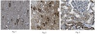
Western Blotting Analysis: A representative lot of MABN893 deteted ABCA1 in lysate of WT mouse brain and demonstrated a loss of signal in lysates of ABCA1 knock out adult mouse brain (Hu et al., 2008. J Lipid Res. 2008 Feb;49(2):386-93.) Immunohistochemistry Analysis: A 1:250 dilution from a representative lot detected ABCA1 in human cerebral cortex, thalamus, and kidney tissues. Produced in collaboration with MAB Institute, Inc.
Biological Information
Immunogen
Linear peptide corresponding to the C-terminus of Mouse ABCA1.
Evaluated by Western Blotting in Calyculin and Okadaic Acid treated NIH-3T3 cell lysate.
Western Blotting Analysis: A 1 µg/mL diultion of this antibody detected ABCA1 in 10 µg of Calyculin and Okadaic Acid treated NIH-3T3 cell lysate.
Usage Statement
Unless otherwise stated in our catalog or other company documentation accompanying the product(s), our products are intended for research use only and are not to be used for any other purpose, which includes but is not limited to, unauthorized commercial uses, in vitro diagnostic uses, ex vivo or in vivo therapeutic uses or any type of consumption or application to humans or animals.
Storage and Shipping Information
Storage Conditions
Stable for 1 year at -20°C from date of receipt. Handling Recommendations: Upon receipt and prior to removing the cap, centrifuge the vial and gently mix the solution. Aliquot into microcentrifuge tubes and store at -20°C. Avoid repeated freeze/thaw cycles, which may damage IgG and affect product performance.
To investigate the interaction of ATP-binding cassette transporter A1 (ABCA1) with calmodulin in relation to its calpain-mediated degradation because many calpain substrates bind calmodulin to regulate cellular functions.The activity of ABCA1 is regulated through proteolysis by calpain. An immunoprecipitation and glutathione S-transferase pull-down assay revealed that ABCA1 directly binds calmodulin in a Ca(2+)-dependent manner. The cytoplasmic loop of ABCA1 contains a typical calmodulin binding sequence of 1-5-8-14 motifs (1245 to 1257 amino acids). The peptide of this region showed binding to calmodulin, and deletion of the 1-5-8-14 motif abolished this interaction. This motif is located near the ABCA1 Pro-Glu-Ser-Thr sequence, and the presence of calmodulin/Ca(2+) protected the peptides from proteolysis by calpain. The knockdown of calmodulin by a specific small and interfering RNA increased the degradation of ABCA1 and decreased ABCA1 protein and apolipoprotein A-I-mediated lipid release. Surprisingly, calmodulin inhibitor W7 increased calmodulin binding to ABCA1 and protected it from calpain-mediated degradation, consistent with our previous finding that this compound increased apolipoprotein A-I-mediated cell cholesterol release.Calmodulin directly binds and stabilizes ABCA1 in the presence of Ca(2+) and increases the generation of high-density lipoprotein.
Biogenesis of HDL by SAA is dependent on ABCA1 in the liver in vivo. Hu, W; Abe-Dohmae, S; Tsujita, M; Iwamoto, N; Ogikubo, O; Otsuka, T; Kumon, Y; Yokoyama, S Journal of lipid research
49
386-93
2008
Serum amyloid A (SAA) was markedly increased in the plasma and in the liver upon acute inflammation induced by intraperitoneal injection of lipopolysaccharide (LPS) in mice, and SAA in the plasma was exclusively associated with HDL. In contrast, no HDL was present in the plasma and only a small amount of SAA was found in the VLDL/LDL fraction (d < 1.063 g/ml) after the induction of inflammation in ABCA1-knockout (KO) mice, although SAA increased in the liver. Primary hepatocytes isolated from LPS-treated wild-type (WT) and ABCA1-KO mice both secreted SAA into the medium. SAA secreted from WT hepatocytes was associated with HDL, whereas SAA from ABCA1-KO hepatocytes was recovered in the fraction that was >1.21 g/ml. The behavior of apolipoprotein A-I (apoA-I) was the same as that of SAA in HDL biogenesis by WT and ABCA1-KO mouse hepatocytes. Lipid-free SAA and apoA-I both stabilized ABCA1 and caused cellular lipid release in WT mouse-derived fibroblasts, but not in ABCA1-KO mouse-derived fibroblasts, in vitro when added exogenously. We conclude that both SAA and apoA-I generate HDL largely in hepatocytes only in the presence of ABCA1, likely being secreted in a lipid-free form to interact with cellular ABCA1. In the absence of ABCA1, nonlipidated SAA is seemingly removed rapidly from the extracellular space.