


ST1053 Sigma-AldrichAnti-20S Proteasome Core Subunits Rabbit pAb
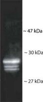

This Anti-20S Proteasome Core Subunits Rabbit pAb is validated for use in Immunoblotting, Immunoprecipitation, Paraffin Sections for the detection of 20S Proteasome Core Subunits.
More>> This Anti-20S Proteasome Core Subunits Rabbit pAb is validated for use in Immunoblotting, Immunoprecipitation, Paraffin Sections for the detection of 20S Proteasome Core Subunits. Less<<Anti-20S Proteasome Core Subunits Rabbit pAb: SDB (Sicherheitsdatenblätter), Analysenzertifikate und Qualitätszertifikate, Dossiers, Broschüren und andere verfügbare Dokumente.
Empfohlene Produkte
Übersicht
| Replacement Information |
|---|
Key Spec Table
| Species Reactivity | Host | Antibody Type |
|---|---|---|
| H, M, Yeast | Rb | Polyclonal Antibody |
Preis & Verfügbarkeit
| Bestellnummer | Verfügbarkeit | Verpackung | St./Pkg. | Preis | Menge | |
|---|---|---|---|---|---|---|
| ST1053-100UL |
|
Kst.-Ampulle | 100 ul |
|
— |

| References |
|---|
| Product Information | |
|---|---|
| Form | Liquid |
| Formulation | Serum diluted in PBS. |
| Positive control | HeLa cells, RSV 3T3 mouse fibroblasts, or yeast whole cell lysates |
| Preservative | ≤0.1% sodium azide |
| Quality Level | MQ100 |
| Biological Information | |
|---|---|
| Immunogen | human erythrocyte proteasomes |
| Immunogen | Human |
| Host | Rabbit |
| Isotype | IgG |
| Species Reactivity |
|
| Antibody Type | Polyclonal Antibody |
| Physicochemical Information |
|---|
| Dimensions |
|---|
| Materials Information |
|---|
| Toxicological Information |
|---|
| Safety Information according to GHS |
|---|
| Safety Information |
|---|
| Product Usage Statements |
|---|
| Packaging Information |
|---|
| Transport Information |
|---|
| Supplemental Information |
|---|
| Specifications |
|---|
| Global Trade Item Number | |
|---|---|
| Bestellnummer | GTIN |
| ST1053-100UL | 04055977224382 |
Documentation
Anti-20S Proteasome Core Subunits Rabbit pAb SDB
| Titel |
|---|
Anti-20S Proteasome Core Subunits Rabbit pAb Analysenzertifikate
| Titel | Chargennummer |
|---|---|
| ST1053 |