Millipore Sigma Vibrant Logo

MABS1253 Anti-O-GlcNAc Antibody, clone RL1

View Products on Sigmaaldrich.com
MABS1253
100 µL  
Price could not be retrieved
Minimum Quantity is a multiple of
Maximum Quantity is
Upon Order Completion More Information
You Saved ()
 
Request Pricing
Limited Availability
In Stock 
Discontinued
Limited Quantities Available
Availability to be confirmed
    Remaining : Will advise
      Remaining : Will advise
      Will advise
      Contact Customer Service
      Contact Customer Service

      Special Offers


      Contact Customer Service

      Overview

      Key Spec Table

      Species ReactivityKey ApplicationsHostFormatAntibody Type
      R, Xn, H, DrABA, EM, ICC, IF, IHC, IP, Inhibition, WBMPurifiedMonoclonal Antibody
      Description
      Catalogue NumberMABS1253
      DescriptionAnti-O-GlcNAc Antibody, clone RL1
      Alternate Names
      • β-GlcNAc
      • β-linked N-acetylglucosamine
      • O-GlcNAc
      • O-linked β-GlcNAc
      Background InformationPosttranslational modification of proteins by -linked N-acetylglucosamine ( -GlcNAc) via the hydroxyl moieties on serine or threonine residues is termed O-linked -GlcNAc or simply O-GlcNAc. O-GlcNAc is one of the most abundant posttranslational modifications within the nucleocytoplasmic compartments of all animals and plants. Unlike other types of protein glycosylations, O-GlcNAc occurs exclusively within the nuclear and cytoplasmic compartments and is generally not further modified to form more elongated structures. In addition, O-GlcNAcylation is a highly dynamic and reversible process. The O-GlcNAc transferase (OGT) attaches O-GlcNAc to proteins at specific serine or threonine residues, while O-GlcNAcase catalyzes the removal/hydrolysis of O-GlcNAc from proteins. In fact, a dynamic interplay between O-GlcNAcylation and serine/threonine phosphorylation plays an important role in regulating cellular signaling. Tau and RNA polymerase II (Pol II) are two well known proteins that undergo modification by O-GlcNAcylation. In Alzheimer s diseased human brains, tau becomes extensively phosphorylated and less O-GlcNAcylated. Similarly, O-GlcNAc is removed and replaced with O-phosphate on the Poly II CTD when the elongation phase of transcription is initiated.
      Product Information
      FormatPurified
      PresentationPurified mouse IgM in buffer containing PBS without preservatives.
      Quality LevelMQ100
      Applications
      ApplicationDetect GlcNAcylated proteins using this mouse monoclonal Anti-O-GlcNAc, clone RL1 , Cat. No. MABS1253, validated for use in Affinity Binding, Electron Microscopy, Immunocytochemistry, Immunofluorescence, Immunohistochemistry, Immunoprecipitation, Inhibition, and Western Blotting.
      Key Applications
      • Affinity Binding Assay
      • Electron Microscopy
      • Immunocytochemistry
      • Immunofluorescence
      • Immunohistochemistry
      • Immunoprecipitation
      • Inhibition
      • Western Blotting
      Application NotesImmunohistochemistry Analysis: A 1:50 dilution from a representative lot detected O-GlcNAcylated cytoplasmic and nuclear pore proteins in rat kidney and spleen tissue sections.

      Affinity Binding Assay: A representative lot of clone RL1 & clone RL2 (Cat. No. MABS157) partially competed against each other for binding immobilized 180 kDa O-GlcNAcylated nuclear envelope protein (Snow, C.M., et al. (1987). J. Cell Biol. 104(5):1143-11560).

      Electron Microscopy Analysis: A representative lot immunolocalized target proteins at the cytoplasmic and/or nucleoplasmic margins of the pore complex with no specific staining of the perinuclear space of isolated rat liver nuclear envelopes (Snow, C.M., et al. (1987). J. Cell Biol. 104(5):1143-11560).

      Immunocytochemistry Analysis: Representative lots stained the nuclear envelope of paraformaldehyde-fixed, Triton X-100-permeabilized HeLa cells, NRK normal rat kidney epithelial cells and isolated rat liver nuclei by fluorescent immunocytochemistry (Yang, L., et al. (1997). J. Cell Biol. 139(5):1077-1087; Byrd, D.A., et al. (1994). J. Cell Biol. 127(6 Pt 1):1515-1526; Snow, C.M., et al. (1987). J. Cell Biol. 104(5):1143-11560).

      Immunofluorescence Analysis: A representative lot stained the nuclear envelope proteins in formaldehyde-fixed, Triton X-100-permeabilzied salivary glands dissected from third-instar Drosophila larvae by fluorescent immunohistochemistry (Goldberg, M., et al. (1998). Mol. Cell. Biol. 18(7):4315-4323).

      Immunofluorescence Analysis: Representative lots stained the nuclear envelope of methanol-fixed xenopus ovary and rat liver cryosections by fluorescent immunohistochemistry (Featherstone, C., et al. (1988). J. Cell Biol. 107(4):1289-1297; Snow, C.M., et al. (1987). J. Cell Biol. 104(5):1143-11560).

      Immunoprecipitation Analysis: A representative lot immunoprecipitated several nuclear envelope proteins in salt-washed nuclear envelope preparations from rat liver and NRK normal rat kidney epithelial cells. Galactosylation of GlcNAc on nuclear envelope proteins by galactosyltransferase treatment significantly inhibited the immunoadsorption of these nuclear pore complex glycoproteins by clone RL1 (Snow, C.M., et al. (1987). J. Cell Biol. 104(5):1143-11560; Holt, G.D., et al. (1987). J. Cell Biol. 104(5):1157-1164).

      Inhibition Analysis: A representative lot, when injected in the vegetal hemisphere of xenopus oocyte cytoplasm, inhibited nucleoplasmin nuclear import in a dose-dependent manner without affecting myoglobin nuclear import or RNA export (Featherstone, C., et al. (1988). J. Cell Biol. 107(4):1289-1297).

      Western Blotting Analysis: Representative lots detected several nuclear envelope proteins in Xenopus oocyte nucleus extract and in salt-washed rat liver nuclear envelope preparations, including target bands of 210, 180, 145, 100, 63, 58, 54, and 45 kDa. GlcNAc removal by beta-N-acetylglucosaminidase treatment greatly reduced the detection of these nuclear pore complex glycoproteins by clone RL1 (Byrd, D.A., et al. (1994). J. Cell Biol. 127(6 Pt 1):1515-1526; Featherstone, C., et al. (1988). J. Cell Biol. 107(4):1289-1297; Snow, C.M., et al. (1987). J. Cell Biol. 104(5):1143-11560; Holt, G.D., et al. (1987). J. Cell Biol. 104(5):1157-1164).
      Biological Information
      ImmunogenNuclear pore complex-lamina fraction isolated from rat liver nuclear envelopes.
      CloneRL1
      ConcentrationPlease refer to lot specific datasheet.
      HostMouse
      SpecificityBoth clone RL1 & clone RL2 (Cat. No. MABS157) target O-linked N-acetylglucosamine (GlcNAc) modified (GlcNAcylated) proteins. GlcNAc removal by beta-N-acetylglucosaminidase treatment or galactosylation of GlcNAc on target proteins by galactosyltransferase treatment greatly reduced the immunoreactivity of clone RL1 & RL2 toward their target proteins. The binding affinity of RL1 and RL2 toward a given GlcNAcylated protein is dependent on both O-GlcNAc and the adjacent core protein structure (Snow, C.M., et al. (1987). J. Cell Biol. 104(5):1143-11560; Holt, G.D., et al. (1987). J. Cell Biol. 104(5):1157-1164).
      IsotypeIgMκ
      Species Reactivity
      • Rat
      • Xenopus
      • Human
      • Drosophila
      Species Reactivity NoteDrosophila, Human, Rat, Xenopus. Target structure is not species-specific.
      Antibody TypeMonoclonal Antibody
      Molecular WeightVariable, depending on the size(s) of the O-GlcNAcylated protein(s). ~210/180/145/100/63/58/54/45 kDa reproted (Snow, C.M., et al. (1987). J. Cell Biol. 104(5):1143-11560; Holt, G.D., et al. (1987). J. Cell Biol. 104(5):1157-1164). Uncharacterized band(s) may appear in some lysates.
      Product Usage Statements
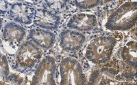
      Quality AssuranceEvaluated by Immunohistochemistry in rat colon tissue.

      Immunohistochemistry Analysis: A 1:50 dilution of this antibody detected O-GlcNAcylated cytoplasmic and nuclear pore proteins in rat colon tissue sections.
      Usage Statement
      • Unless otherwise stated in our catalog or other company documentation accompanying the product(s), our products are intended for research use only and are not to be used for any other purpose, which includes but is not limited to, unauthorized commercial uses, in vitro diagnostic uses, ex vivo or in vivo therapeutic uses or any type of consumption or application to humans or animals.
      Storage and Shipping Information
      Storage ConditionsStable for 1 year at -20°C from date of receipt.
      Handling Recommendations: Upon receipt and prior to removing the cap, centrifuge the vial and gently mix the solution. Aliquot into microcentrifuge tubes and store at -20°C. Avoid repeated freeze
      Packaging Information
      Material Size100 µL
      Global Trade Item Number
      Catalogue Number GTIN
      MABS1253 04054839055577