Millipore Sigma Vibrant Logo

ST1069 PhosphoDetect™ Anti-Paxillin (pSer¹⁷⁸) Rabbit pAb

View Products on Sigmaaldrich.com
ST1069
  
Retrieving price...
Price could not be retrieved
Minimum Quantity is a multiple of
Maximum Quantity is
Upon Order Completion More Information
You Saved ()
 
Request Pricing
Limited Availability
In Stock 
Discontinued
Limited Quantities Available
Availability to be confirmed
    Remaining : Will advise
      Remaining : Will advise
      Will advise
      Contact Customer Service
      Contact Customer Service


      Contact Customer Service

      Overview

      Replacement Information

      Key Spec Table

      Species ReactivityHostAntibody Type
      H, MRbPolyclonal Antibody
      Description
      OverviewRecognizes the ~68 kDa paxillin protein phosphorylated at Ser178.
      Catalogue NumberST1069
      Brand Family Calbiochem®
      Application Data
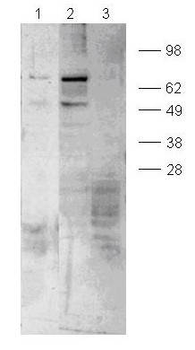
      Detection of mouse paxilin phosphorylated at Ser178, by immunoblotting. Samples: Whole cell lysate (~2.4 x 10⁵ cells/lane) from NH3T3 cells serum-starved for 16 h and grown in suspension (lane 1) NIH3T3 cells plated on fibronectin (50 µg/ml for 1 h, lane 2), and NIH3T3 cells plated on fibronectin and treated with lamda phosphotase (lane 3). Primary antibody: Phospho-Dtect™ Anti-Paxilin (pSer178) Rabbit pAb (Cat. No. ST1069) (1:1000). Detection: chemiluminescence.
      References
      ReferencesBellis, S.L., et al. 1997. Biochem. J. 325, 375.
      Schaller, M.D. 2001. Oncogene 20, 6459.
      Huang, C., et al. 2003. Nature 424, 219.
      Product Information
      FormLiquid
      FormulationIn Tris-citrate/phosphate, pH 7.0-8.0.
      Positive controlNIH3T3 cells plated on fibronectin
      Preservative≤0.1% sodium azide
      Quality LevelMQ100
      Applications
      Key Applications Immunoblotting (Western Blotting)
      Application NotesImmunoblotting (1:1000-1:5000)
      Application CommentsDoes not recognize unphosphorylated paxillin. Antibody should be titrated for optimal results in individual systems.
      Biological Information
      Immunogena synthetic phosphopeptide corresponding to amino acids surrounding the Ser¹⁷⁸ phosphorylation site of human paxillin
      ImmunogenHuman
      HostRabbit
      IsotypeIgG
      Species Reactivity
      • Human
      • Mouse
      Antibody TypePolyclonal Antibody
      Concentration Label Please refer to vial label for lot-specific concentration
      Physicochemical Information
      Dimensions
      Materials Information
      Toxicological Information
      Safety Information according to GHS
      Safety Information
      Product Usage Statements
      Storage and Shipping Information
      Ship Code Blue Ice Only
      Toxicity Highly Toxic
      Storage +2°C to +8°C
      Do not freeze Ok to freeze
      Special InstructionsFollowing initial use aliquot and freeze (-20°C) for long-term storage. Avoid freeze/thaw cylces.
      Packaging Information
      Transport Information
      Supplemental Information
      Specifications
      Global Trade Item Number
      Catalogue Number GTIN
      ST1069 0

      Documentation

      PhosphoDetect™ Anti-Paxillin (pSer¹⁷⁸) Rabbit pAb Certificates of Analysis

      TitleLot Number
      ST1069

      References

      Reference overview
      Bellis, S.L., et al. 1997. Biochem. J. 325, 375.
      Schaller, M.D. 2001. Oncogene 20, 6459.
      Huang, C., et al. 2003. Nature 424, 219.
      Data Sheet

      Note that this data sheet is not lot-specific and is representative of the current specifications for this product. Please consult the vial label and the certificate of analysis for information on specific lots. Also note that shipping conditions may differ from storage conditions.

      Revision18-October-2007 RFH
      ApplicationImmunoblotting (1:1000-1:5000)
      Application Data
      Detection of mouse paxilin phosphorylated at Ser178, by immunoblotting. Samples: Whole cell lysate (~2.4 x 10⁵ cells/lane) from NH3T3 cells serum-starved for 16 h and grown in suspension (lane 1) NIH3T3 cells plated on fibronectin (50 µg/ml for 1 h, lane 2), and NIH3T3 cells plated on fibronectin and treated with lamda phosphotase (lane 3). Primary antibody: Phospho-Dtect™ Anti-Paxilin (pSer178) Rabbit pAb (Cat. No. ST1069) (1:1000). Detection: chemiluminescence.
      DescriptionImmunoaffinity purified rabbit polyclonal antibody. Recognizes the ~68 kDa paxillin protein phosphorylated at Ser178.
      BackgroundPaxillin is a focal adhesion adaptor protein associated with th establishment of focal adhesions, cell signaling via integrins, and cell motility. The N-terminus of paxillin contains a proline-rich region for SH3 and SH2 domain binding. The N-terminus also contains five copies of the LD motif for association of actin binding proteins. The C-terminus contains LIM domains and the focal adhesion targeting sequence. Paxillin is believed to function as an adaptor protein that facilitates assembly of multi-protein complexes and coordinates signaling via integrins. Tyrosine and serine phosphorylation of paxillin is associated with the establishment of cytoskeletal membrane attachments. Phosphorylation by JNK at Ser178 is necessary for normal cell motility.
      HostRabbit
      Immunogen speciesHuman
      Immunogena synthetic phosphopeptide corresponding to amino acids surrounding the Ser¹⁷⁸ phosphorylation site of human paxillin
      IsotypeIgG
      Specieshuman, mouse
      Positive controlNIH3T3 cells plated on fibronectin
      FormLiquid
      FormulationIn Tris-citrate/phosphate, pH 7.0-8.0.
      Concentration Label Please refer to vial label for lot-specific concentration
      Preservative≤0.1% sodium azide
      CommentsDoes not recognize unphosphorylated paxillin. Antibody should be titrated for optimal results in individual systems.
      Storage +2°C to +8°C
      Do Not Freeze Ok to freeze
      Special InstructionsFollowing initial use aliquot and freeze (-20°C) for long-term storage. Avoid freeze/thaw cylces.
      Toxicity Highly Toxic
      ReferencesBellis, S.L., et al. 1997. Biochem. J. 325, 375.
      Schaller, M.D. 2001. Oncogene 20, 6459.
      Huang, C., et al. 2003. Nature 424, 219.