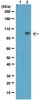
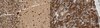
Millipore Sigma Vibrant Logo

DP11 Anti-Adenovirus 2 E1A Mouse mAb (M73)

DP11
  
Retrieving price...
Price could not be retrieved
Minimum Quantity is a multiple of
Maximum Quantity is
Upon Order Completion More Information
You Saved ()
 
Request Pricing
Limited Availability
In Stock 
Discontinued
Limited Quantities Available
Availability to be confirmed
    Remaining : Will advise
      Remaining : Will advise
      Will advise
      Contact Customer Service
      Contact Customer Service


      Contact Customer Service

      Overview

      Replacement Information

      Key Spec Table

      Species ReactivityHostAntibody Type
      Adenovirus Infected CellsMMonoclonal Antibody
      Description
      Overview

      This product has been discontinued.


      Recognizes the adenovirus 2 E1A and 5 E1A proteins, immunoprecipitating 3 proteins at ~30-35 kDa, as well as associated proteins such as Rb, p53, and cyclins.

      Catalogue NumberDP11
      Brand Family Calbiochem®
      References
      ReferencesWhyte, P., et al. 1998. Nature 334, 124.
      Ziff, E. et al. 1985. Cell 40, 705.
      Houweling, A., et al. 1980. Virology 105, 537.
      Berk, A.J., et al. 1979. Cell 17, 935.
      Jones, N. and Shenk, T. 1979. Proc. Natl. Acad. Sci. USA 76, 3665.
      Shiroki, K., et al. 1979. Virology 95, 127.
      Gallimore, P.H., et al. 1974. J. Mol. Biol. 89, 49.
      Graham, F.L., et al. 1974. Cold Spring Harbor Symp. Quant. Biol. 39, 637.
      Product Information
      FormLiquid
      FormulationIn 50 mM sodium phosphate buffer, 0.2% gelatin.
      Negative controlHS27 cells
      Positive controlHEK293 cells or adenovirus-infected tissue
      Preservative≤0.1% sodium azide
      Quality LevelMQ100
      Applications
      Application ReferencesImmunoblotting Stiewe T., et al. 2000. Cancer Res. 60, 3957. Nishihara, A., et al. 1999. J. Biol. Chem. 274, 28716. Sanchez-Prieto, R., et al. 1999. Nat. Med. 5, 1076. van den Berg, A., et al. 2005. Respir. Res. 6, 111. Immunocytochemistry van den Berg, A., et al. 2005. Respir. Res. 6, 111. Immunofluoresecence Harlow, E., et al. 1986. Mol. Cell. Biol. 6, 1579. Harlow, E., et al. 1985. J. Virol. 3, 533. Original Clone Harlow, E., et al. 1985. J. Virol. 3, 533. Paraffin Sections Nemunaitis, H., et al. 2000 Cancer Res. 60, 6359.
      Key Applications Frozen Sections
      Immunoblotting (Western Blotting)
      Immunocytochemistry
      Immunofluorescence
      Immunoprecipitation
      Paraffin Sections
      Application NotesImmunofluorescence (2-5 µg/ml, see application references)
      Immunoprecipitation (1-2 µg antibody/mg total protein)
      Immunoblotting (1 µg/ml; see application references)
      Immunocytochemistry (see application references)
      Frozen Sections (2-4 µg/ml)
      Paraffin Sections (2-4 µg/ml; heat pre-treatment required; see application references)
      Application CommentsImmunoprecipitates the adenovirus 2 and 5 E1A proteins (3 bands at ~30-50 kDa) as well as associated proteins. Staining of formalin-fixed, paraffin-embedded tissues requires boiling tissue sections in 10 mM citrate buffer, pH 6.0, for 10-20 min followed by cooling at RT for 20 min. Antibody should be titrated for optimal results in individual systems.
      Biological Information
      Immunogenadenovirus 2 E1A protein
      CloneM73
      HostMouse
      IsotypeIgG2a
      Species Reactivity
      • Adenovirus Infected Cells
      Antibody TypeMonoclonal Antibody
      Concentration Label Please refer to vial label for lot-specific concentration
      Physicochemical Information
      Dimensions
      Materials Information
      Toxicological Information
      Safety Information according to GHS
      Safety Information
      Product Usage Statements
      Storage and Shipping Information
      Ship Code Blue Ice Only
      Toxicity Standard Handling
      Storage +2°C to +8°C
      Do not freeze Yes
      Special InstructionsFollowing initial use, aliquot and freeze (-20°C) for long-term storage.
      Packaging Information
      Transport Information
      Supplemental Information
      Specifications
      Global Trade Item Number
      Catalogue Number GTIN
      DP11 0

      Documentation

      Anti-Adenovirus 2 E1A Mouse mAb (M73) Certificates of Analysis

      TitleLot Number
      DP11

      References

      Reference overview
      Whyte, P., et al. 1998. Nature 334, 124.
      Ziff, E. et al. 1985. Cell 40, 705.
      Houweling, A., et al. 1980. Virology 105, 537.
      Berk, A.J., et al. 1979. Cell 17, 935.
      Jones, N. and Shenk, T. 1979. Proc. Natl. Acad. Sci. USA 76, 3665.
      Shiroki, K., et al. 1979. Virology 95, 127.
      Gallimore, P.H., et al. 1974. J. Mol. Biol. 89, 49.
      Graham, F.L., et al. 1974. Cold Spring Harbor Symp. Quant. Biol. 39, 637.

      Citations

      Title
    • Ricardo Sanchez-Prieto, et al. (1999) An Association Between Viral Genes and Human Oncogenic Alterations: The Adenovirus E1A Induces the Ewing Tumor Fusion Transcript EWS-FLI1. Nature Medicine 5, 1076-1079.
    • Data Sheet

      Note that this data sheet is not lot-specific and is representative of the current specifications for this product. Please consult the vial label and the certificate of analysis for information on specific lots. Also note that shipping conditions may differ from storage conditions.

      Revision30-January-2009 JSW
      ApplicationImmunofluorescence (2-5 µg/ml, see application references)
      Immunoprecipitation (1-2 µg antibody/mg total protein)
      Immunoblotting (1 µg/ml; see application references)
      Immunocytochemistry (see application references)
      Frozen Sections (2-4 µg/ml)
      Paraffin Sections (2-4 µg/ml; heat pre-treatment required; see application references)
      DescriptionProtein A purified mouse monoclonal antibody generated by immunizing Balb/c mice with the specified immunogen and fusing splenocytes with NS-1 mouse myeloma cells (see application references). Recognizes adenovirus-infected cells and tissue.
      BackgroundThe early region (E1) of the adenovirus genome, responsible for transforming activity, is localized within the leftmost 11% of the viral genome and consists of two transcriptional units, E1A and E1B. Region E1A is sufficient for partial transformation and immortalization of primary cells, whereas the E1B function is normally required for complete transformation. In addition to their essential role in transformation, E1A gene products are necessary for normal levels of transcription of the other early regions of the adenovirus genome during productive infection, and are able to either activate or repress the transcription of specific cellular genes. E1A oncogene proteins form specific complexes with cellular proteins including the anti-oncogene retinoblastoma susceptibility gene product, p105. The consequence of this interaction is inhibition of the cell cycle arresting function of p105.
      HostMouse
      Immunogenadenovirus 2 E1A protein
      CloneM73
      IsotypeIgG2a
      Speciesadenovirus infected cells
      Positive controlHEK293 cells or adenovirus-infected tissue
      Negative controlHS27 cells
      FormLiquid
      FormulationIn 50 mM sodium phosphate buffer, 0.2% gelatin.
      Concentration Label Please refer to vial label for lot-specific concentration
      Preservative≤0.1% sodium azide
      CommentsImmunoprecipitates the adenovirus 2 and 5 E1A proteins (3 bands at ~30-50 kDa) as well as associated proteins. Staining of formalin-fixed, paraffin-embedded tissues requires boiling tissue sections in 10 mM citrate buffer, pH 6.0, for 10-20 min followed by cooling at RT for 20 min. Antibody should be titrated for optimal results in individual systems.
      Storage +2°C to +8°C
      Do Not Freeze Yes
      Special InstructionsFollowing initial use, aliquot and freeze (-20°C) for long-term storage.
      Toxicity Standard Handling
      ReferencesWhyte, P., et al. 1998. Nature 334, 124.
      Ziff, E. et al. 1985. Cell 40, 705.
      Houweling, A., et al. 1980. Virology 105, 537.
      Berk, A.J., et al. 1979. Cell 17, 935.
      Jones, N. and Shenk, T. 1979. Proc. Natl. Acad. Sci. USA 76, 3665.
      Shiroki, K., et al. 1979. Virology 95, 127.
      Gallimore, P.H., et al. 1974. J. Mol. Biol. 89, 49.
      Graham, F.L., et al. 1974. Cold Spring Harbor Symp. Quant. Biol. 39, 637.
      Citation
    • Ricardo Sanchez-Prieto, et al. (1999) An Association Between Viral Genes and Human Oncogenic Alterations: The Adenovirus E1A Induces the Ewing Tumor Fusion Transcript EWS-FLI1. Nature Medicine 5, 1076-1079.
    • Application referencesImmunoblotting Stiewe T., et al. 2000. Cancer Res. 60, 3957. Nishihara, A., et al. 1999. J. Biol. Chem. 274, 28716. Sanchez-Prieto, R., et al. 1999. Nat. Med. 5, 1076. van den Berg, A., et al. 2005. Respir. Res. 6, 111. Immunocytochemistry van den Berg, A., et al. 2005. Respir. Res. 6, 111. Immunofluoresecence Harlow, E., et al. 1986. Mol. Cell. Biol. 6, 1579. Harlow, E., et al. 1985. J. Virol. 3, 533. Original Clone Harlow, E., et al. 1985. J. Virol. 3, 533. Paraffin Sections Nemunaitis, H., et al. 2000 Cancer Res. 60, 6359.